Contact Us Careers
Lung Cancer

The patient with sarcoma developed lung metastasis and underwent NKT treatment for two years, resulting in these changes

时间:2026-04-27 人气:
Ms. G is suffering from a relatively rare type of soft tissue sarcoma (Ewing's sarcoma). Sarcomais different from cancer, but both belong to malignant tumors. The primary difference between sarcoma and cancer lies in the origin of the tissue. Cancer is a malignant tumor that originates from epithelial tissue, while sarcoma is a malignant tumor that originates from connective tissue, muscle, and other tissues, hence the term "sarcoma".
Sarcoma is more common in adolescents, but its incidence rate is low, whereas cancer is more prevalent in adults. Due to its subtle early symptoms, most patients with cancer are prone to metastasis and diffusion when discovered, and recurrence is also common in the middle and late stages.
In terms of metastasis patterns, cancer is more commonly metastasized to lymph nodes through the lymphatic system, while sarcoma is more likely to metastasize to the liver, lungs, and bones through hematogenous spread, as these areas have richer blood supply. Compared to cancer, sarcoma is rarer, and related research and treatment are also less extensive.
During the treatment of Ms. G for more than two years, there was no significant progression in her condition, and even some lesions showed shrinkage, which is very meaningful for the treatment of this type of sarcoma originating from mesenchymal tissue.

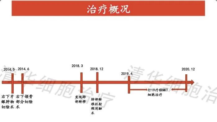
The patient discovered a painless mass in the right lower gum in March 2014, measuring approximately 2*1cm. On May 17th, an outpatient surgical excision was performed. Pathology indicated a spindle cell tumor in the right lower posterior region of the tooth, involving bone tissue. One lymph node was removed and no metastasis was found, suggesting a need for external consultation.

On June 3, 2014, the Fudan University Affiliated Cancer Hospital provided a consultation: (right lower posterior region of the tooth) soft tissue sarcoma. Based on the EWSR1 translocation, it is inclined to be mesenchymal chondrosarcoma or extrabony Ewing's sarcoma.
On June 25, 2014, a partial mandibulectomy was performed under general anesthesia, and no cancerous tissue was found in the intraoperative pathology.
On March 13, 2018, chest CT: multiple nodules in both lungs, with possible metastasis.
On April 9, 2018, pathology (left lung biopsy) revealed soft tissue sarcoma, consistent with soft tissue sarcoma metastasis in the right lower posterior region of the jaw.
On June 13, 2018, CT showed multiple metastases in both lungs, with a larger one in the left lower lung measuring approximately 1.2 cm.
On August 13, 2018, CT showed multiple nodules in both lungs, with possible metastasis.
On December 5, 2018, percutaneous radiofrequency ablation of the right lung tumor was performed.
On January 16, 2019, chest CT: changes after radiofrequency ablation of the nodule in the right middle lung lobe, with the nodule slightly smaller than before. Multiple metastases in the remaining two lungs remained similar to before.
On March 31, 2019, CT scan showed multiple small nodules in both lungs, with calcification visible in some of the lesions, the largest being approximately 1.9cm. There were scattered few streaky shadows in both lungs.
On April 2, 2019, NKT cell therapy was initiated, with a regimen of one course per month (due to COVID-19 in February and March 2020, no reinfusion was performed). During this period, four imaging assessments were stable, and as of today (June 21, 2021), 21 courses have been completed.
Through timely, continuous, and systematic individualized NKT cell immunotherapy, along with seven treatment follow-ups.
In terms of imaging:
After the patient started NKT cell immunotherapy, the largest metastatic lesion in the lungs had shrunk from 1.5cm*1.2cm (as of March 2019) to completely disappear by January 2021. The nodule in the left lower lung had slightly shrunk compared to before. Compared to the previous year, not only had the patient's condition not progressed, but many positive changes had also occurred.

Chest CT: nodules with patchy shadows in the right lung middle lobe, slightly smaller in March 2019 compared to January, slightly reduced in July 2019 compared to March, no significant change from July to December 2019, reduced in June 2020 compared to March 2020, unclear in January 2021, disappeared compared to previous images.
The disappearance of lung metastases may be attributed to the effect of radiofrequency ablation. However, various physical treatments, including radiofrequency ablation, typically have immediate effects, meaning they take effect quickly but also "lose effectiveness" quickly.
Therefore, observing the rate of reduction/disappearance of metastatic lesions and calculating the duration of maintaining the effect are important criteria for measuring the efficacy of cellular immunotherapy.
In this case, the lung metastases gradually disappeared over a period of one year, and no new lesions appeared during the two-year period. It must be said that NKT immunotherapy played a significant role!

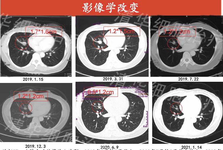
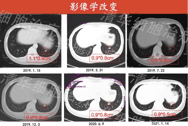
Chest CT: nodules in the left lower lobe of the lung with uneven density, calcification visible within, slightly reduced in size from March 2019 compared to January 2019, no significant change from March 2019 to June 2020, and slightly smaller in January 2021 compared to June 2020.

Chest CT: low-density nodules visible near the capsule of the right lobe of the liver, filling-like changes after enhancement, likely hemangioma, no significant change from January 2019 to January 2021.
Regarding tumor markers and quality of life:

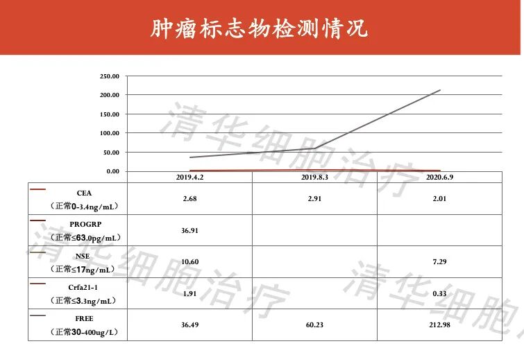
From April 2019 to August 2021, all tumor markers tested were within the normal range and remained stable. After NKT treatment, with the reduction of tumor burden, the patient's quality of life improved significantly, with a weight gain of 1-3kg, good sleep and appetite, and a quality of life score of 92 points.

Assessment results:

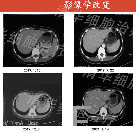
Ms. G underwent 21 courses of NKT cell immunotherapy, with almost no visible maximum metastatic lesion in the lungs, and the remaining smaller metastatic lesions either shrank or remained unchanged. No new signs of disease progression were observed in other organs. Her progression-free survival (PFS) reached 27 months, and she continues to receive treatment and follow-up.

NKT Classic Case Review

Click on the image

to view